Xerra Imaging Platform
Fully automated CFT workflow to advance discoveries in biological and drug research
Contact UsXerra™: EMIT Imaging's Flagship Platform
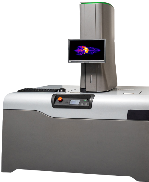
XerraTM, is EMIT Imaging’s flagship preclinical imaging platform available for purchase. Xerra is designed to advance discoveries in biological and drug research and fully automates the CFT workflow, providing 3D images with high resolution and sensitivity. This enables researchers to easily run samples in-house, and obtain unmatched visualization of drug distribution and protein expression in whole-animals. See what you’re missing compared to standard 2D in vivo fluorescence imaging techniques and utilize CFT to discover more.
Xerra provides:
- Automated CFT workflow
- Anatomical, RGB white-light images registered with fluorescence images
- Capable of Multiplexing
- 5 magnifications: 20-55 µm pixel resolution
- 6 excitation lasers: 470 to 780 nm
- 7 emission filters: 500 to 850 nm
How CFT Works
Step 1: Preparation
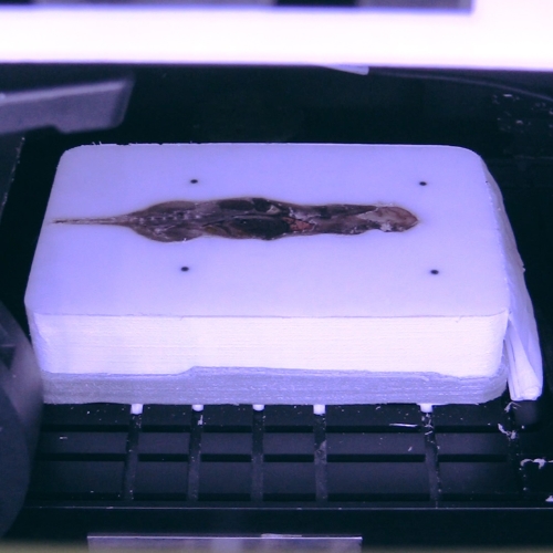
Step 2: Image + Section
Step 3: Output
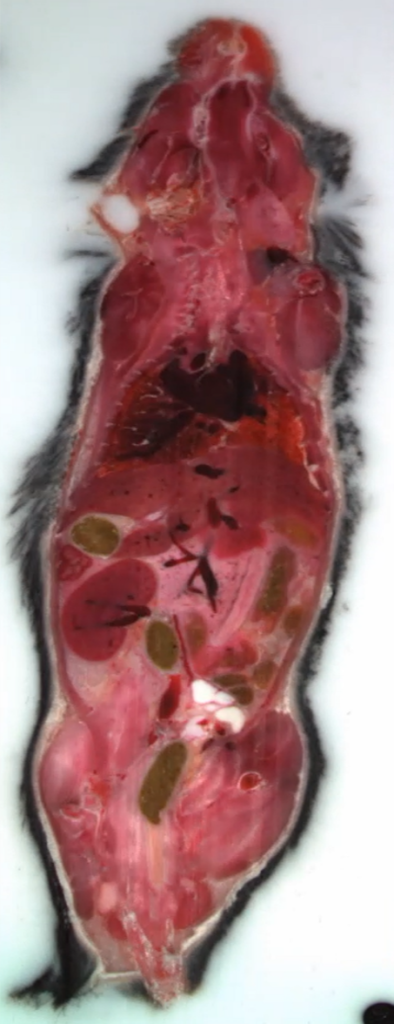

3D image stacks are generated. White light and fluorescence data can be overlaid.
Research Advantages
High Resolution:
High, near-cellular resolution down to 20 µm
High Sensitivity:
Nanomolar sensitivity, comparable to nuclear medicine
High Throughput:
Several samples in the same block = large volume of tissue imaged
Complementary:
Fits into any experiment, complementing tissue and radiology imaging
Simplified:
No fixation, perfusion, clearing or radiolabeling required
Multiplexing:
6 lasers and 7 filters for multiplexed applications
Specifications
High Resolution
20 µm to 55 µm isotropic voxels depending on FOV
True 3D
Reconstruct whole body & large tissue data from white light and fluorescence images
Automated
Leverage built-in, turn-key workflows for image acquisition
- Obtain automated reconstruction of 2D slices into 3D
- Anatomical, RGB white-light images registered with high sensitivity fluorescence imaging, capable of multiplexing
Simplified
Sample preparation is simple and does not require fixation, perfusion, clearing, or radiolabeling
- Sac to image in as little as 4 hours. Frozen samples can be stored for weeks before imaging as long as they are not blocked in OCT
- Xerra is compatible with most existing fluorophores
- Xerra requires no infrastructure changes for install
Multiplexing
Xerra has 6 excitation lasers ranging from 470 to 780 nm and 7 emission filters ranging from 500 to 850 nm
Quantification
CFT has advantages for quantification and EMIT Imaging fully characterizes fluorophores and optimizes laser power exposure time, attenuating conditions, and subsurface signal analysis to improve quantification
- Signal is surface-weighted
- Signal is linear
6 Excitation Lasers
470-780 nm
7 Emission Filters
500-850 nm
Record Multiple Exposures
Recording at variable exposure times (5, 50, 500, 1500, 25000 milliseconds)
Max: 24 cm x 14 cm
Min: 8 cm x 5 cm
Working Volume(s)
FOV A: Pixel – 20 µm, Block Size – 8x6x4 cm, Sample – Tissue
FOV B: Pixel – 30 µm, Block Size – 10x8x5 cm, Sample – 1 Mouse
FOV C: Pixel – 35 µm, Block Size – 14x11x6 cm, Sample – 3 Mice
FOV D: Pixel – 45 µm, Block Size – 18x14x8 cm, Sample – 4 Mice
FOV E: Pixel – 55 µm, Block Size – 24x14x10 cm, Sample – 5 Mice or 1 Rat
Section Thickness
20 μm to 50 μm
Display
Quality Analysis (QA)
Manual Section Collection
Yes
Refrigerated chamber, -20°. Auto defrost functionality
Image Analysis and Quantification
Compatible with Various Software Packages
Data Management
WIFI connectivity or USB A and C data ports