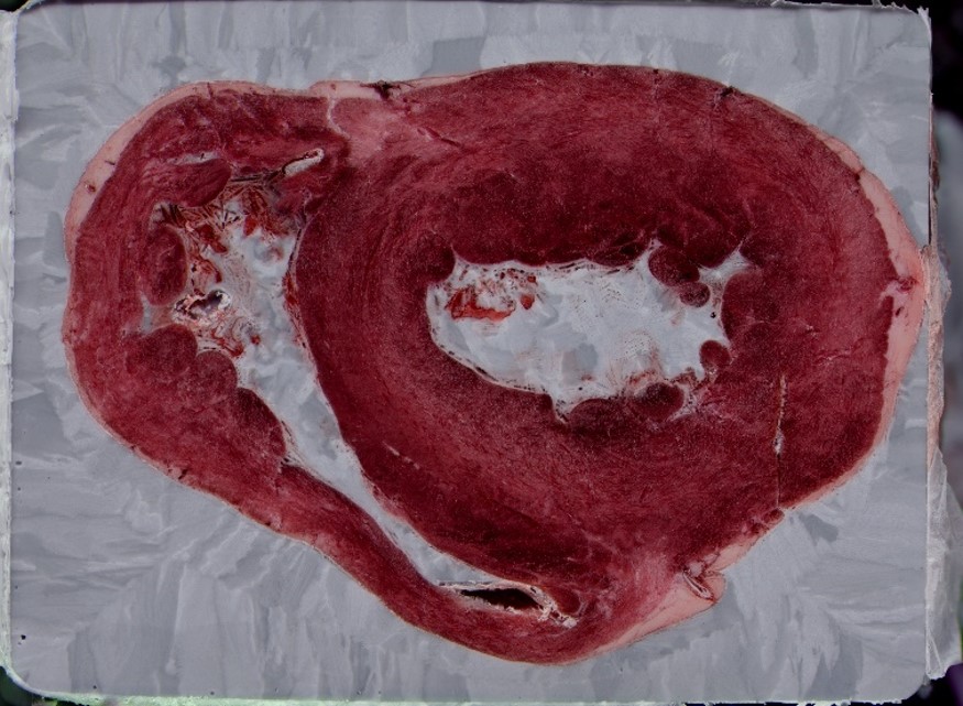
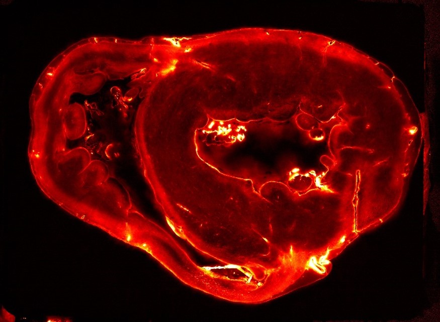
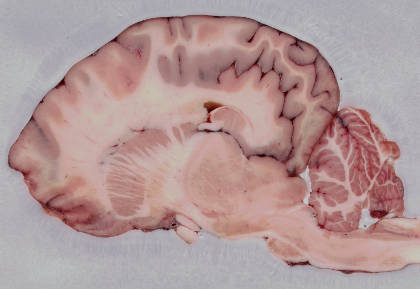
Cryo-fluorescence tomography (CFT) is an ex vivo, volumetric tissue imaging technique used to monitor the pharmacokinetics and pharmacodynamics (PK/PD) of drugs and delivery vehicles with high-resolution and sensitivity. Images of the anatomy (white light) are acquired alongside fluorescence images that are displayed in a 3D visualization of the drug and/or delivery vehicle biodistribution.
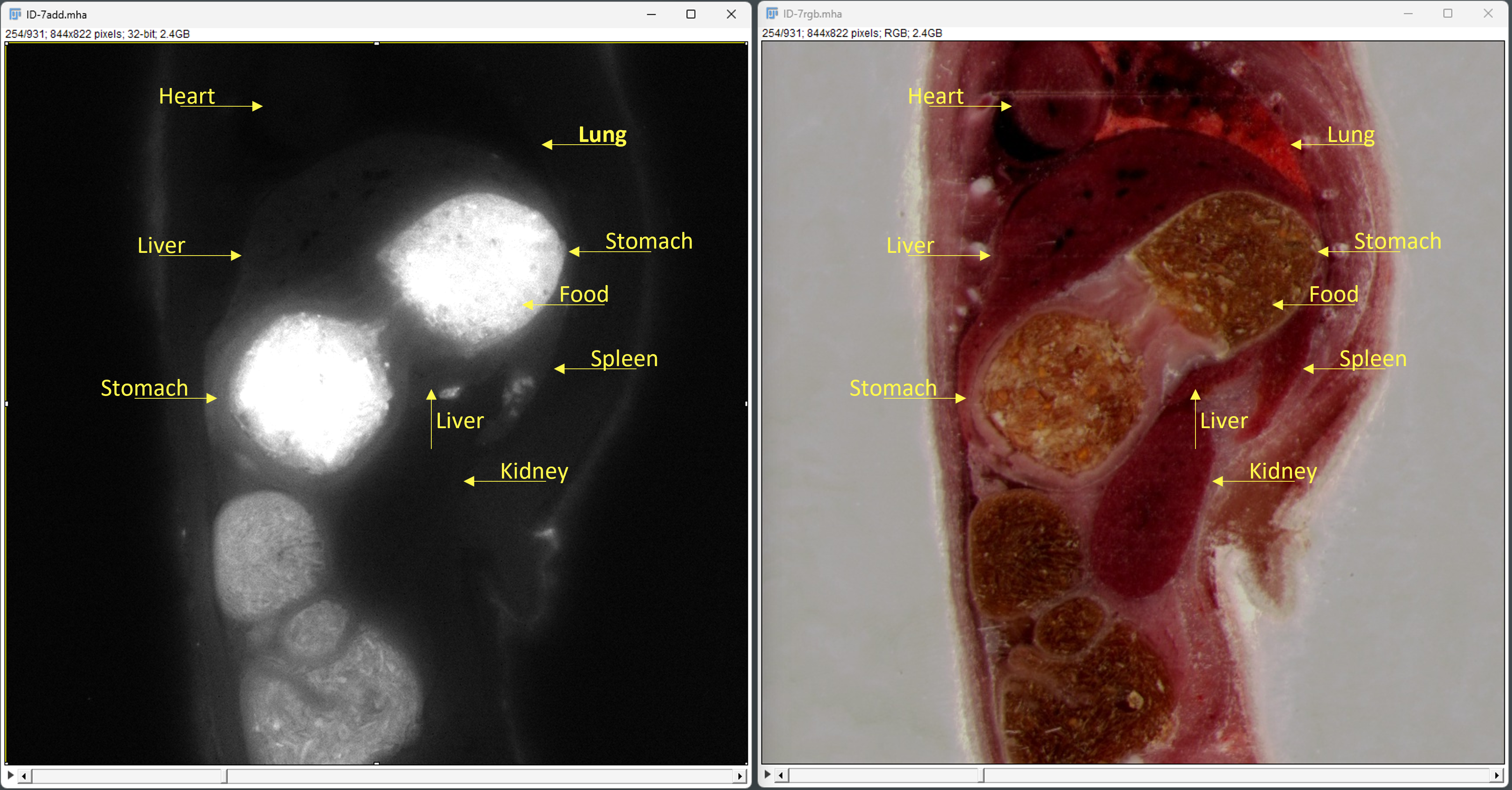
Findings: CFT uniquely achieves the required combination of resolution and sensitivity in a whole animal.
Details:
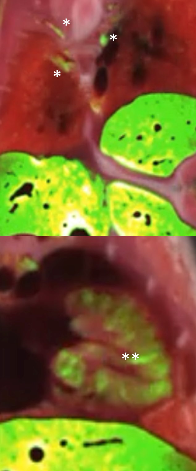
Findings: CFT was used to visualize labeled cell intake into tissue.
Details:
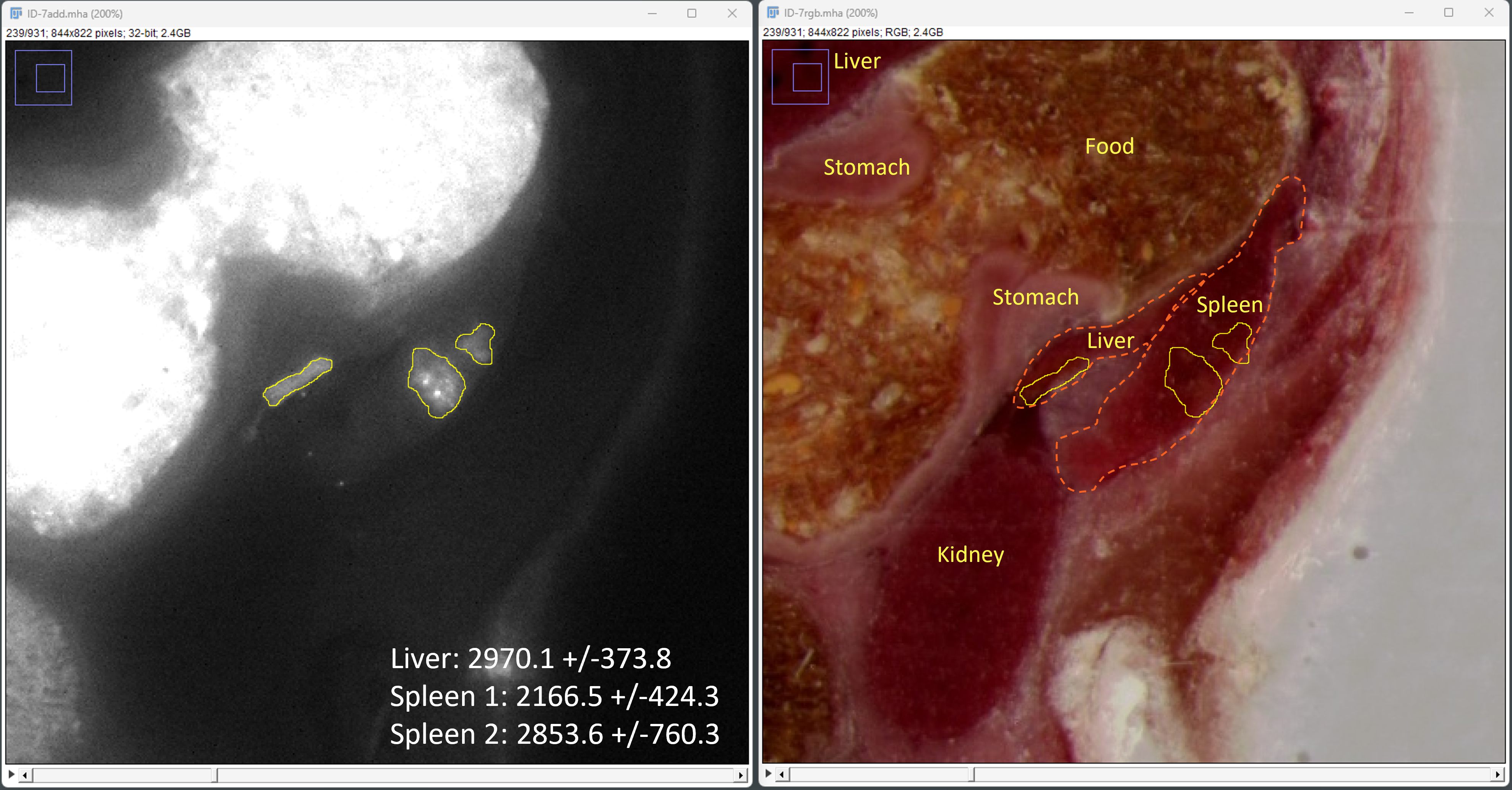
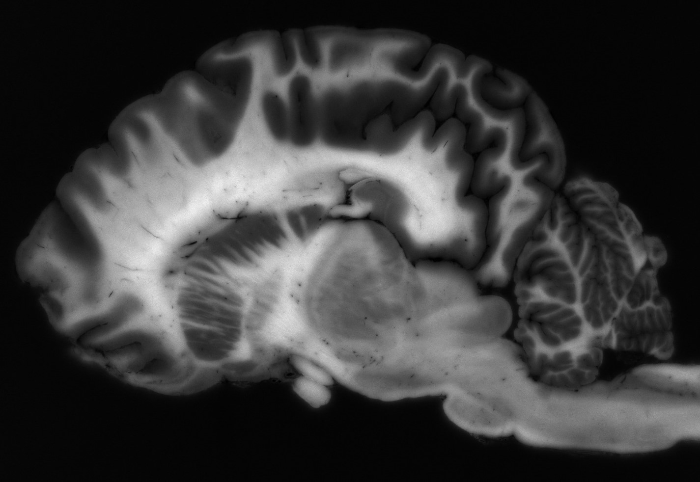
Because Xerra works in large formats, CFT has strong applications for anatomical RGB white-light and fluorescence imaging in large animal tissue, including NHP, Bovine, Porcine, and more.
| Cookie | Duration | Description |
|---|---|---|
| cookielawinfo-checkbox-analytics | 11 months | This cookie is set by GDPR Cookie Consent plugin. The cookie is used to store the user consent for the cookies in the category "Analytics". |
| cookielawinfo-checkbox-functional | 11 months | The cookie is set by GDPR cookie consent to record the user consent for the cookies in the category "Functional". |
| cookielawinfo-checkbox-necessary | 11 months | This cookie is set by GDPR Cookie Consent plugin. The cookies is used to store the user consent for the cookies in the category "Necessary". |
| cookielawinfo-checkbox-others | 11 months | This cookie is set by GDPR Cookie Consent plugin. The cookie is used to store the user consent for the cookies in the category "Other. |
| cookielawinfo-checkbox-performance | 11 months | This cookie is set by GDPR Cookie Consent plugin. The cookie is used to store the user consent for the cookies in the category "Performance". |
| viewed_cookie_policy | 11 months | The cookie is set by the GDPR Cookie Consent plugin and is used to store whether or not user has consented to the use of cookies. It does not store any personal data. |