


Cryo-Fluorescence Tomography (CFT) and EMIT Imaging’s flagship platform Xerra™ are powerful techniques for oncology and immunotherapy applications. CFT provides anatomical & 3D fluorescence images to help researchers to discover more when studying tumor models including microenvironments, tumor heterogeneity, metastatic spread, and expression of specific biomarkers.
For metastatic tumor progression, CFT detects metastatic disease and provides high resolution 3D molecular data of tumor burden and spread, not typically visualized in other image modalities. In addition, CFT is used to evaluate tumor metabolism and cell biology in response to genetic manipulations, pharmacologic agents, and cancer chemotherapy drugs.
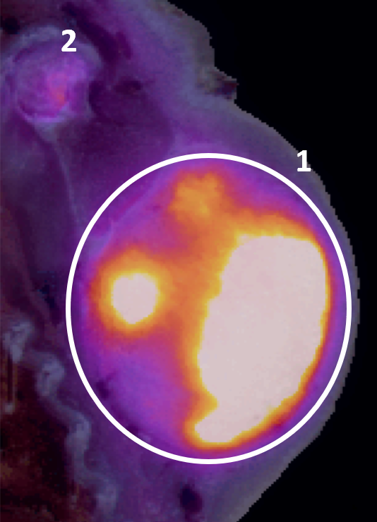
Findings: Data confirms drug preferential uptake in the tumor with minimal uptake in other organs/tissues
Details:
Copyright 2023 Ratio Therapeutics Inc., used with permission
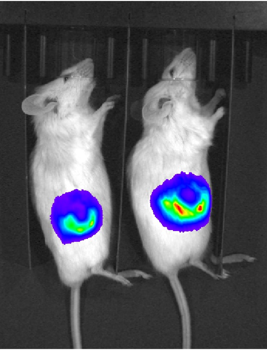
Findings: CFT shows high-resolution, 3D co-localization of tumor cells and drugs, complementing and expanding 2D in vivo imaging
Details
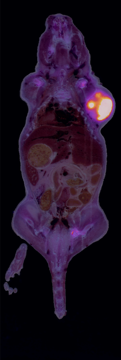
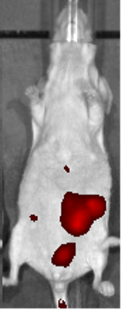
Findings: CFT image shows the primary tumor extent and heterogeneity as well as the metastatic lesions in the lungs
Details:
DsRed mice provided by John Ronald, Robarts Research Institute
| Cookie | Duration | Description |
|---|---|---|
| cookielawinfo-checkbox-analytics | 11 months | This cookie is set by GDPR Cookie Consent plugin. The cookie is used to store the user consent for the cookies in the category "Analytics". |
| cookielawinfo-checkbox-functional | 11 months | The cookie is set by GDPR cookie consent to record the user consent for the cookies in the category "Functional". |
| cookielawinfo-checkbox-necessary | 11 months | This cookie is set by GDPR Cookie Consent plugin. The cookies is used to store the user consent for the cookies in the category "Necessary". |
| cookielawinfo-checkbox-others | 11 months | This cookie is set by GDPR Cookie Consent plugin. The cookie is used to store the user consent for the cookies in the category "Other. |
| cookielawinfo-checkbox-performance | 11 months | This cookie is set by GDPR Cookie Consent plugin. The cookie is used to store the user consent for the cookies in the category "Performance". |
| viewed_cookie_policy | 11 months | The cookie is set by the GDPR Cookie Consent plugin and is used to store whether or not user has consented to the use of cookies. It does not store any personal data. |